Article

Protected Freshwater Ecosystem with Incessant Cyanobacterial Blooming Awaiting a Resolution

1
Department of Biology and Ecology, Faculty of Sciences, University of Novi Sad, Trg Dositeja Obradovića 3, 21000 Novi Sad, Serbia
2
Department of Aquaculture, Szent István University, Páter Károly u. 1, 2100 Gödöllő, Hungary
3
Faculty of Science, Institute of Biology and Ecology, University of Kragujevac, Radoja Domanovića 12, 34 000 Kragujevac, Serbia
4
Biochemistry, Faculty of Science and Engineering, Åbo Akademi University, Tykistökatu 6 A, 20520 Turku, Finland
5
Finnish Environment Institute, Marine Research Centre, Agnes Sjöbergin katu 2, 00790 Helsinki, Finland
*
Author to whom correspondence should be addressed.
Water 2020, 12(1), 129; https://doi.org/10.3390/w12010129
Submission received: 27 November 2019 / Revised: 27 December 2019 / Accepted: 29 December 2019 / Published: 31 December 2019
(This article belongs to the Special Issue Advancing Knowledge on Cyanobacterial Blooms in Freshwaters)

Abstract

:
For 50 years persistent cyanobacterial blooms have been observed in Lake Ludoš (Serbia), a wetland area of international significance listed as a Ramsar site. Cyanobacteria and cyanotoxins can affect many organisms, including valuable flora and fauna, such as rare and endangered bird species living or visiting the lake. The aim was to carry out monitoring, estimate the current status of the lake, and discuss potential resolutions. Results obtained showed: (a) the poor chemical state of the lake; (b) the presence of potentially toxic (genera Dolichospermum, Microcystis, Planktothrix, Chroococcus, Oscillatoria, Woronichinia and dominant species Limnothrix redekei and Pseudanabaena limnetica) and invasive cyanobacterial species Raphidiopsis raciborskii; (c) the detection of microcystin (MC) and saxitoxin (STX) coding genes in biomass samples; (d) the detection of several microcystin variants (MC-LR, MC-dmLR, MC-RR, MC-dmRR, MC-LF) in water samples; (e) histopathological alterations in fish liver, kidney and gills. The potential health risk to all organisms in the ecosystem and the ecosystem itself is thus still real and present. Although there is still no resolution in sight, urgent remediation measures are needed to alleviate the incessant cyanobacterial problem in Lake Ludoš to break this ecosystem out of the perpetual state of limbo in which it has been trapped for quite some time.

1. Introduction

In the very north of Serbia there is an old and unusual lake, Lake Ludoš, with beautiful open water landscapes surrounded by reeds, wetlands and steppe. The environment is rich in many plant and animals species. European pond turtles, various amphibians, otters, moles, rabbits, foxes and roe deer have found their home there. What makes Lake Ludoš especially famous, and validates the name originating from the Hungarian word “lud” meaning goose, is that there are more than 200 bird species, including rare and endangered species, nesting or resting during their migration. Because of all this, Lake Ludoš is recognized and protected as a special nature reserve on the list of Ramsar sites.
One part of the lake is often visited by fishermen, but their catch mostly consists of the very resilient and adaptable Prussian carp (Carassius gibelio (Block, 1782)) which are quite small in size. This may be related to the fact that the water of Lake Ludoš has an intense green color throughout the year caused by cyanobacteria (e.g., [1]). Their extensive growth and blooming causes many problems in freshwater ecosystems, including this one. Cyanobacteria can produce cyanotoxins that affect other organisms, including valuable flora and fauna, especially aqueous organisms such as fish [2]. Cyanobacteria also present a threat to humans, such as fishermen, who may be exposed to cyanotoxins through contaminated food, inhalation and direct contact [3,4]. In addition to human health, the health of this important ecosystem is also jeopardized. Furthermore, this “disease” could also be transmissible, since there is a possibility that water birds visiting the lake during their migration path can disseminate toxic cyanobacteria [5].
Lake Ludoš is only one of many aquatic ecosystems in Serbia where cyanobacteria are present and blooming [6]. What sets this ecosystem apart from many others is that it has been known for perpetual blooming in the last 50 years. Previous research has shown that the lake is in a poor ecological state which leads to the question of whether the protection of this natural habitat in a bad ecological state is justified. The cyanobacterial problem, which can potentially affect every living being in the proximity of this ecosystem, has also been preserved as measures to improve the water quality have not been undertaken on the lake [5]. The problem of cyanobacteria in Lake Ludoš has been addressed during our previous research when potentially toxic cyanobacterial species, cyanotoxins in water, macrophytes and fish tissues were detected, as well as histological alterations and DNA damage in fish tissue (see [5]). Six years later further monitoring was carried out in order to estimate the current state of this ecosystem. Therefore, several investigative steps have been taken: monitoring of physical and chemical parameters of the lake; assessment of qualitative and quantitative analyses of cyanobacteria; the first survey of the cyanotoxin coding genes; determination of cyanotoxins in water and fish; analysis of histopathology of different fish organs; and discussion of potential health risks and resolutions.
Freshwater ecosystems throughout the world have similar problems in connection to cyanobacterial blooming and cyanotoxin production. Recent publication of a global geographical and historical overview of cyanotoxin distribution demonstrated the presence of well-known cyanotoxins in each continent (including 520 lakes) and their harmful consequences on human health [4]. Hence, this issue is of global concern. The present investigation aims to assist in making appropriate decisions and measures for the remediation of not only this, but many other old and rapidly aging and protected lakes.

2. Materials and Methods

2.1. Sampling Site and Sampling of Water and Fish

Lake Ludoš (Figure 1) is a one of the few preserved shallow lakes in the region. It has a maximum depth of 2.25 m, is 4.5 km long, and it represents a remnant of the Pannonian Sea. In most places the depth does not exceed 1 m, and it may be frozen for more than three months a year. It is located in the north part of Serbia, near the city of Subotica. The lake and the associated wetland ecosystem is highly valued due to the great biological diversity, and as such the area is classified among wetlands of international significance. The quality of the lake’s water is of great importance for the preservation of the flora and fauna connected to this marshland ecosystem.
The lake is supplied with water from aquifers and Kereš River. However, in the northern part, Lake Ludoš receives water from the canal Palić-Ludoš which is the recipient of wastewaters from the Palić settlement. Water treatment of these wastewaters is still inadequate and the canal water is characterized by a high level of organic pollution, high concentrations of salt and very high nutrient concentrations. The inflow of untreated and partially purified waters in Lake Ludoš contributes to the deterioration of the water quality and the increase of the sludge quantity [7,8,9,10,11].
Water samples were collected from the surface water layer within the littoral zone (pier next to the visitor center) (46.103207 N, 19.821360 E) and from the center of the lake (46.102159 N, 19.821149 E) in March, May, July and September of 2018. Samples of Prussian carp were collected from the center of the lake before and after the summer (March and September 2018) with gillnets of various mesh sizes and a standard electrofishing device.

2.2. Analyses of Physical and Chemical Parameters

Multi-parameter WTW probes were used for carrying out in situ measurements in the field and the following physical and chemical parameters were determined: temperature, pH, conductivity, O2 concentration and O2 saturation. TSS (total suspended solids), TOC (total organic carbon), NO3, detergents, COD (chemical oxygen demand) and BOD (biological oxygen demand) were measured in the laboratory conditions with a Pastel Ultraviolet (UV) Secomam.

2.3. Qualitative and Quantitative Analyses of Cyanobacteria

The phytoplankton samples for the cyanobacterial qualitative analysis were collected by sweeping a plankton net (netframe 25 cm ø, net mesh 23 μm). All samples were immediately preserved in a Lugol solution. Taxonomic identifications of cyanobacteria were made according to several taxonomic keys [12,13,14,15] and were done under a light microscope Motic BA310 using a Bresser (9MP) digital camera and Micro Cam Lab software. For the quantitative analysis of phytoplankton, the 15 L of water was collected by sweeping a plankton net at the depth of 0.3 m in March, while in May, July and September only 200 mL of water were collected directly from the lake as a result of high bloom density. The quantitative analysis was made by using the Utermöhl method [16] under a Motic AE 2000 inverted microscope. The phytoplankton individuals were sedimented and cyanobacteria quantified on the chamber (i.e., transects) with an inverted microscope at different magnifications depending on their size (100×, 400×) and expressed as the number of cells per mL.

2.4. Cyanotoxin Coding Gene Analyses

2.4.1. Samples—Reference Strains for Polymerase Chain Reaction (PCR) Analysis

Reference strains were obtained from Pasteur Culture Collection (PCC), National Institute for Environmental Studies Microbial Culture Collection (NIES), Australian National Algae Culture Collection (CS), and Finnish Environment Institute (SYKE). They consisted of:
  • microcystin (MC) producers: PCC7820 (Microcystis aeruginosa), NIES-107 (Microcystis wesenbergii);
  • cylindrospermopsin (CYN) producers: CS-505, CS-506 (Cylindrospermopsis raciborskii), SYKE-966 (Anabaena lapponica);
  • saxitoxin (STX) producers: CS-337/01, CS-537/13 (Dolichospermum circinale);
  • anatoxin-a (ATX) producer: ANA123 (Dolichospermum circinale).

2.4.2. DNA Extraction

Depending on the bloom density, 30–400 mL of water samples were filtered (pore size 2–3 µm), and filtride was freeze-dried. Approximately 10 mg of freeze-dried biomass was used for DNA extraction from reference strains. Genomic DNA from biomass of the reference strains and filtrides was extracted with the DNeasy Plant Mini Kit (QIAGEN, Hilden, Germany) according to manufacturer’s instructions, with minimal modifications for the extraction from filtrides (double amount of Buffer AP1, RNase A and Buffer P3 was added to fully suspend the samples). During the initial steps of extraction, samples were homogenized using zirconia/silica disruption beads (0.5 mm) and by vortexing for 1 min. The quality was assessed spectrophotometrically (NanoDrop ND-1000, Thermo Scientific, Waltham, MA, USA), where A260/A280 ratio varied between 1.22 and 2.04.

2.4.3. Qualitative PCR

Qualitative PCR was run to analyze samples for the presence of MC (mcyE), CYN (cyrJ), STX (sxtA, sxtG, sxtS) and ATX (anaC) synthetase genes. PCR reaction mixtures were prepared in a total volume of 20 µL containing 1× Phire Reaction Buffer, 0.4 µL Phire II HotStart polymerase (Thermo Scientific), 0.2 mM deoxyribonucleotide triphosphates (dNTPs) (Thermo Scientific), 0.5 µL forward and reversed primers (Table 1), 2 µL of template and sterile deionized water. PCRs were run on a C1000 Touch Thermal Cycler (Bio-Rad, Helsinki, Finland) according to the following protocols: initial denaturation for 30 s at 98 °C; 40 cycles of 5 s at 98 °C, 5 s at 61 °C (HEPF, HEPR, sxtA1480_R stxA855_R), or 62 °C (cyrJ_F, cyrJ_R, sxtG432_F, sxtG928_R, sxtS205_F, sxtS566_R), or 52 °C (anaC-genF, anaC-genR) and 10 s at 72 °C; and a final extension of 1 min at 72 °C. To examine the potential inhibition of PCRs, an exogenous amplification control template was prepared containing 1 µL:1 µL (reference:sample). Following strains were used as a reference in the control template: PCC7820 for mcyE, CS-506 for cyrJ, CS-537/13 for sxtA, sxtG, sxtS, and ANA123 for anaC. Visualization of PCR products was performed on a 1.5% Top Vision agarose gel (Thermo Scientific) dyed with SYBR® Safe DNA gel stain. The observed bands were documented on Gel Doc™ XR (Bio-Rad) using Quantity One software (v. 4.6.9).

2.5. Cyanotoxin Analyses

2.5.1. Preparation of Water Samples for Liquid Chromatography–Tandem Mass Spectrometry (LC–MS/MS)

Depending on the bloom density, 30–100 mL of water samples were filtered (pore size 2–3 µm). Biomass on the filter was then freeze-dried. Afterwards, filtrides were placed in glass tubes and the toxin was extracted with 3 mL of 75% MeOH and ultrasonication. The extracts were centrifuged for 10 min at 10,000× g and two 1 mL aliquots of the supernatant were evaporated to dryness (50 °C nitrogen flow) in glass tubes. For MC analysis the sample was redissolved in 75% MeOH in 200 µL, and for CYN analysis the sample was redissolved in 200 µL H2O. The samples were then filtered (0.2 µm GHP ACRODISC 13 Pall Life Sciences, Ann Arbot, MS, USA) into inserts and were ready for liquid chromatography–tandem mass spectrometry (LC–MS/MS) analysis.
The extracellular MC content was concentrated by solid-phase extraction (SPE) on Waters Oasis HLB (30 mg). The samples eluted with 5 mL 90% MeOH were placed into glass tubes, evaporated using nitrogen flow and redissolved in 200 µL of 75% MeOH. Subsequently, they were filtered (0.2 µm GHPACRODISC 13 Pall Life Sciences) into inserts and were ready for LC–MS/MS analysis.

2.5.2. Preparation of Fish Tissue Samples for LC–MS/MS

Prussian carp from Lake Ludoš was analysed for MCs in fish liver, gills, kidney, intestine, gonads (testis and ovaries), spleen, and muscle samples by LC–MS/MS. A total of 30 individuals (TL: 16.63 ± 2.24 cm, SL: 14.58 ± 1.68 cm, 18 female/12 male) were collected during two separate sampling surveys—spring (March) and autumn (September) of 2018. Different fish tissues were separately homogenized and freeze-dried. Samples of the same organ of all the individuals were pooled together. Before further preparation, several fish tissues were spiked in order to test the preparation method. Freeze-dried fish tissue samples (100 mg) were placed into glass tubes and 5 mL of 75% MeOH was added for extraction of cyanotoxins. Homogenization was performed on ice for 30 second, and the samples were then ultrasonicated in a bath sonicator for 15 min and further extracted with a probe sonicator. Samples were then centrifuged for 10 min at 10,000× g followed by an addition of 1 mL of hexane to 2 mL of the supernatants obtained. The hexane (lipid) layer was removed using glass pipettes and the remaining samples were evaporated (50 °C nitrogen flow) in glass tubes. Finally, samples were redissolved in 300 µL 75% MeOH and filtered (0.2 µm GHPACRODISC 13 Pall Life Sciences) into inserts. The fish tissue samples were then ready for LC-MS/MS analysis.

2.5.3. LC–MS/MS

Toxin analyses were performed by LC–MS/MS [22]. The analytical targets consisted of nine MC variants (MC-dmRR, MC-RR, MC-dmYR, MC-YR, MC-dmLR, MC-LR, MC-LY, MC-LW and MC-LF) and CYN.

2.6. Analyses of Fish Histology

Captured Prussian carp from Lake Ludoš used for cyanotoxin detection were also used for histological analyses. Liver, kidney, gill, intestine, spleen, gonad and muscle samples were dissected from each fish and fixed in 4% formaldehyde. Additionally, 6 individuals of common carp (Cyprinus carpio L.) (TL: 18.35 ± 6.24 cm, 3 females/3 males) were obtained from the Department of Aquaculture, Szent István University, Hungary. These fish were kept in a recirculation system (Sentimento Kft., Hungary), under a 12 h light/12 h dark cycle at 24 ± 0.2 °C and served as a control group in this study.
After fixation of at least three days, samples were processed by standard histological procedure. Gill and muscle samples were decalcified beforehand. For tissue processing, samples were dehydrated in graded series of ethanol, cleared in xylene and subsequently embedded in paraffin wax blocks. Three five-µm-thin sections per tissue per individual were cut and placed onto glass slides, and stained with haematoxylin and eosin (H&E) dyes. Sections were examined under a Nikon Eclipse 600 microscope and photographed using a QImaging Micro Publisher 3.0 digital camera.

3. Results

3.1. Physical and Chemical Parameters of Water Samples

The recent investigations of Lake Ludoš during 2018 corroborated the continuously poor chemical state of the lake. pH levels, saturation with O2 as well as electrical conductivity were high during the investigated period (Table 2).

3.2. Presence of Cyanobacterial Species in Water Samples

Most dominant cyanobacterial species were Limnothrix redekei (Van Goor) Meffert and Pseudanabaena limnetica (Lemmermann) Komárek (Table 3, Figure 2). Furthermore, both Microcystis species, Microcystis aeruginosa (Kützing) Kützing and Microcystis wesenbergii (Komárek) Komárek, were numerous during the whole investigated period. At the end of the summer, an invasive Raphidiopsis raciborskii (Woloszynska) Aguilera, Berrendero Gómez, Kastovsky, Echenique and Salerno (basionym Cylindrospermopsis raciborskii (Woloszynska) Seenayya and Subba Raju) also started to occur. Usually, more cells per mL were found in the pier samples compared to the center samples; however, the same species were present in both sampling sites.

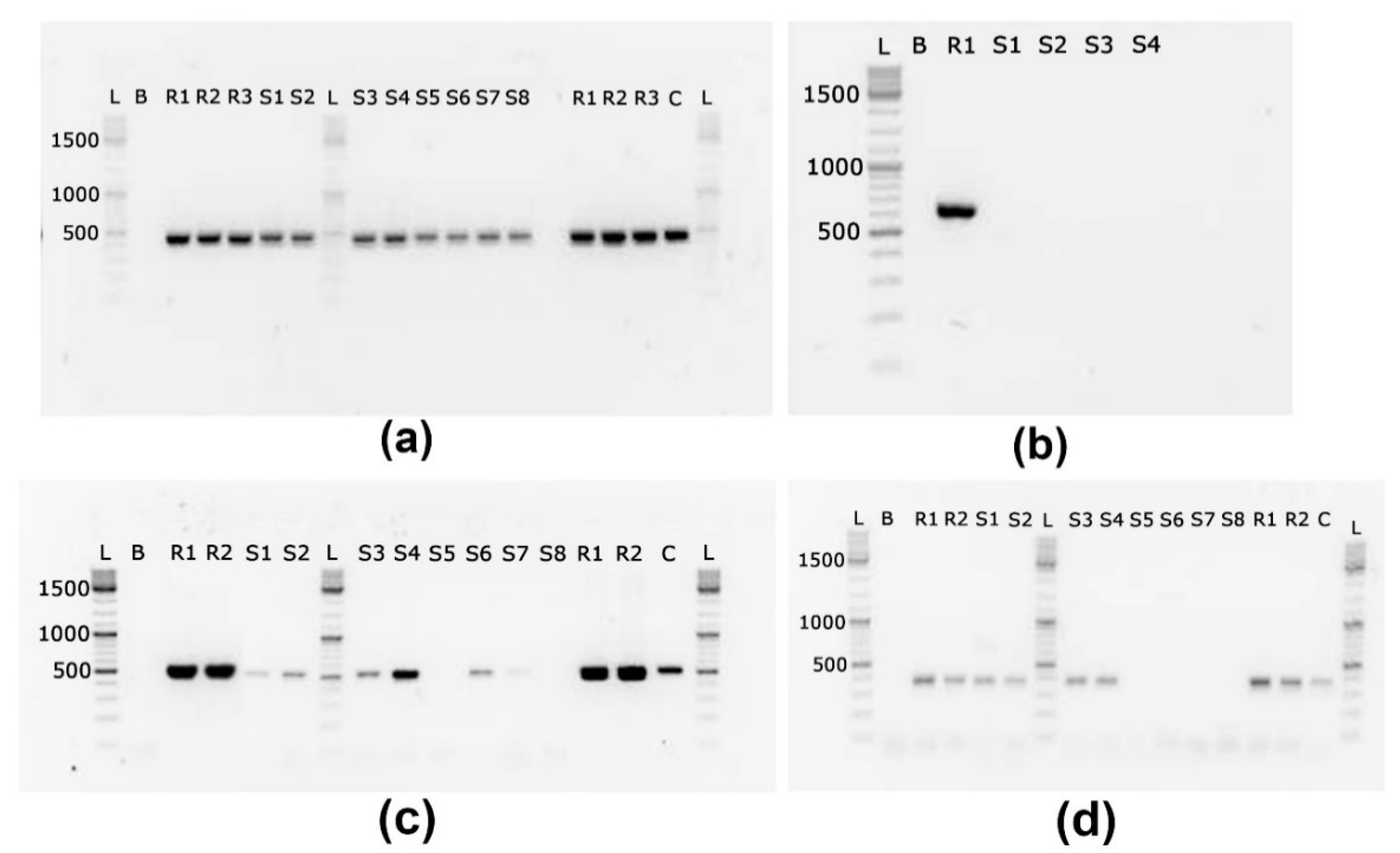
3.3. Presence of Cyanotoxin Coding Genes in Biomass Samples

Biomass samples were tested for the presence of cyanotoxin coding genes, including MCs, STX, CYN and ATX (Table 4).
MC coding gene mcyE (472 bp, Figure 3a) was amplified in all samples. STX coding genes sxtG (519 pb, Figure 3c) and sxtS (382 bp, Figure 3d) were amplified in a total of 6 and 4 samples, respectively. The sxtG gene was observed in all sampling seasons, while sxtS gene was observed only in samples collected in August and September. Saxitoxin coding gene sxtA (648 bp, Figure 3b), CYN coding gene cyrJ, and ATX coding gene anaC were not amplified in this study. Significant inhibition of the PCR reaction was not observed in exogenous amplification control templates.

3.4. Presence of Cyanotoxins in Water and Fish Samples

Biomass and extracellular content of water samples were tested for the presence of cyanotoxins. Several MC variants were noted in the total content (Table 5) including most commonly occurring MC-LR and MC-RR, however, their concentrations were rather low during the whole investigated period. CYN was not detected during the investigated period in Lake Ludoš, even though there were some cyanobacterial species that could potentially produce this cyanotoxin.
Analyses of several fish tissue samples did not show a presence of investigated MC variants in spring nor autumn samples.

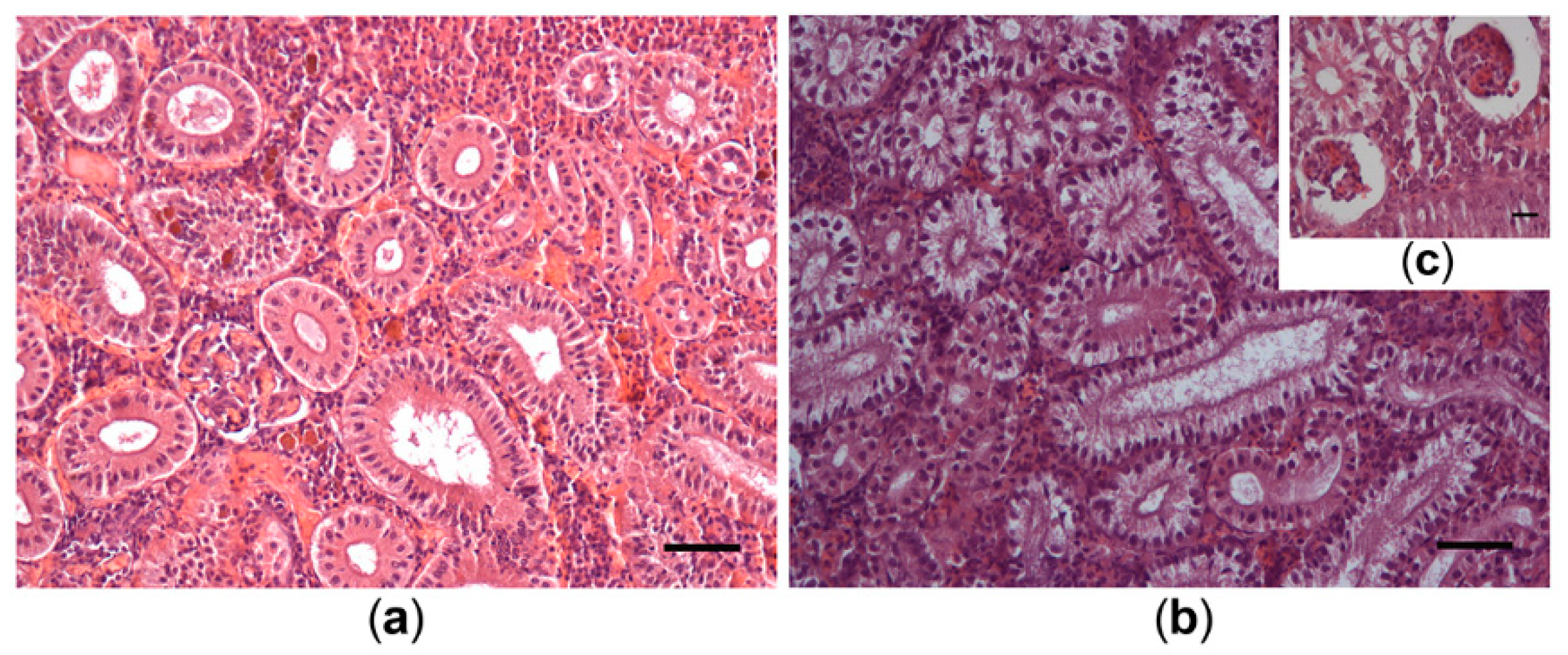
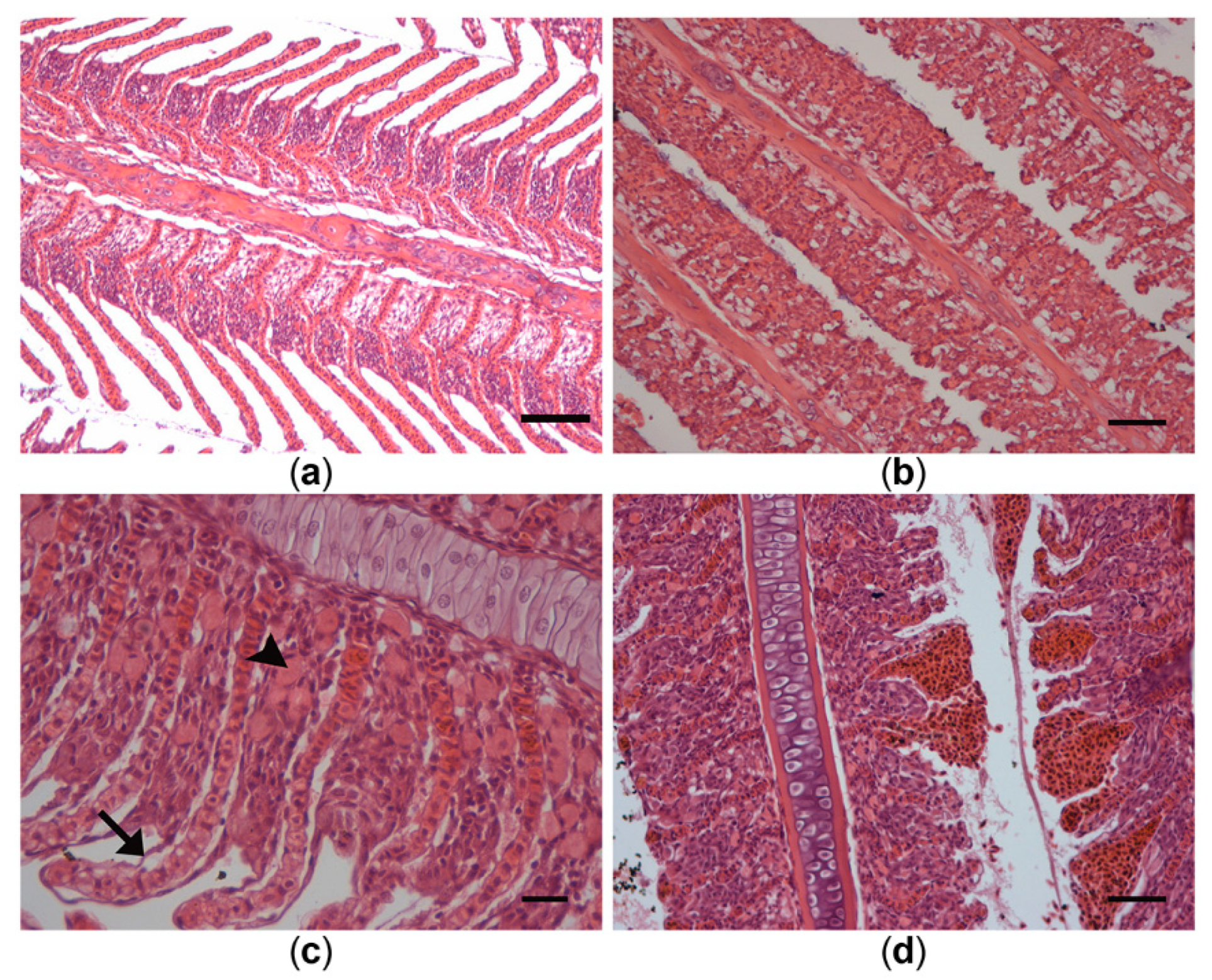
3.5. Histological Alterations in Fish Samples

During both investigated periods, spring (before) and autumn season (during and after the bloom), most affected fish organs were liver, kidneys and gills. Liver samples of individuals from the control group displayed typical organization of the hepatic parenchyma, with cord-like formations of hepatocytes interspaced with sinusoids and radially arranged around blood vessels (Figure 4a). Hepatocytes were polygonal, with a clearly visible cell membrane and large round nuclei with distinguishable nucleoli. In contrast, microscopic examination of fish from Lake Ludoš revealed severe alterations of liver histology which were observed in both March and September sampling groups. Livers of these fish showed changes in architectural structure, with less prominent cord-like organization and sinusoid capillaries no longer clearly distinguishable (Figure 4b). Loss of shape and rounding of hepatocytes was most characteristic in the March group, with large groups of cells displaying ball-or onion-like shape (Figure 4c). Altered hepatocytes typically had darker nuclei with condensed chromatin and no discernable nucleoli. Signs of kariopyknosis, predominantly in the September sampling group, were indicative of necrosis (Figure 4d). Most prominent alterations present in all examined samples were glycogen depletion and vacuolization of hepatocytes. Many cells had a completely clear cytoplasm, which suggest hypervacuolization (Figure 4e).
Renal corpuscles in the kidneys of the control individuals were round in shape and had relatively large glomeruli. The Bowman’s capsule was continuous with thin intercapsular space. Both proximal and distal renal tubules had one layer of columnar epithelial cells with proximal segments having basal nuclei, and distal segments having central nuclei and less intensive cytoplasmic stain (Figure 5a). Even in controls, slight clogging of tubules and slight vacuolization was observed.
Only individuals sampled in March had significant pathological alterations in the kidney. These included degeneration and loss of nephron formation, as well as interstitium structure. Renal corpuscles showed a reduction of glomeruli size, accompanied with dilatations of intercapsular space of the Bowman’s capsule (Figure 5c). In tubules, epithelial cells showed intense vacuolization and in some cases the epithelial layer was separated from the basal lamina (Figure 5b). The number of tubules appeared clogged and in the process of necrosis. Cells in the necrotic area had pyknotic nuclei and displayed signs of cell membrane lysis, such as no discernable boundary between cells. In some fish, necrosis, hyalization of the interstitium and the presence of macrophage aggregates were evident. Certain alterations, such as vacuolization and tearing of the tubular epithelium, were also present in individuals from the September group; however, this was not as frequent and severe compared to the March group.
Gills of the control group showed no pathological changes (Figure 6a). Secondary lamellae regularly lined both sides of the primary lamellae (filament) and were covered with one layer of squamous epithelial cells. Contrarily, individuals from Lake Ludoš in both sampling periods (March and September) had noticeably altered gill structure. Several individuals displayed signs of hyperplasia, as well as hypertrophy of interlamellar cell mass, mainly epithelial and mucous cells. Such swelling of secondary lamellae and proliferation of interepithelial cells has led to a complete fusion of the secondary lamellae, especially noticeable in the September sampling group (Figure 6b). Other observed lesions included epithelial lifting and oedema, accompanied with hypertrophy of interepithelial chloride cells (Figure 6c). In some individuals, endothelial cells of the capillaries showed signs of telangiectasia (aneurysm), along with epithelial rupture and hemorrhage (Figure 6d).
Other examined organs of the bloom-exposed fish in the present study did not display histopathological alterations (not shown). Intestines of all groups showed normal histology, with villi regularly lining the lumen. A single layer of enterocytes with basal nuclei lined the surface of the vili, along with fewer goblet cells. Furthermore, sections of muscle tissue in all groups also showed no structural alterations. Spleen of all examined individuals displayed a normal structure, with aggregates of erythroid and lymphoid cells, surrounding blood vessels. Neither male nor female gonads had histopathological changes. Testes and ovaries had normal structural organization with germline cells present in different stages and numbers, which is dependent on the season and age of the fish.

4. Discussion

4.1. Monitoring of the Water

4.1.1. Physical and Chemical Parameters in Lake Ludoš

The measurements of physical and chemical parameters showed several important findings:
  • the lake water pH values were very high (almost 9), probably as a results of the activity of phytoplankton;
  • O2 saturation showed high values, and even supersaturation during July 2018, likely due to photosynthetic activity of phytoplankton;
  • electrical conductivity was relatively high.
It is assumed that cyanobacteria are more resistant to solute increases compared to other phyla e.g., Chlorophyta [23]. Additionally, conductivity changes lead to decreased zooplankton—predators of phytoplankton, thus it is possible that a reduction in zooplankton would potentiate phytoplankton increase.
Regular monitoring showed similar findings in recent years (2013–2017) [7,8,9,10,11]. pH levels were between 8 and 9, O2 saturation during the year was uneven with high values and supersaturation during summer and sometimes during autumn months, while high values of electric conductivity of the water seemed to be rising in the recent years. Additionally, COD was extremely high, similar to wastewaters, indicating a poor status of the lake and based on the measured parameters it is recommended that the lake should not to be used for any purpose [24]. BOD was uneven during the year, demonstrating the problem of instability of the system. The water of the Lake Ludoš is very rich in nitrogen compounds (mostly organically bound nitrogen), which led to increased biological production. Nitrates were uneven during the year and seemed to depend on the inflow from the Palić-Ludoš canal. Phosphates showed a similar trend as nitrates and their concentration seemed to be on the rise thus contributing to the deterioration of the water quality. Sediment analysis suggested rich deposits of nutrients which will contribute to the continual hypertrophic state of the lake [7,8,9,10,11].
The status of surface waters in terms of general quality can be shown by the Serbian Water Quality Index (SWQI). SWQI is based on 10 quality parameters (temperature, pH, conductivity, O2 saturation, BOD for 5 days, suspended matter, total nitrogen oxides, orthophosphates, ammonia ions, coliform bacteria) that are aggregated into a composite indicator of the quality of surface waters, leading them to one index number [24]. SWQI of the water from Lake Ludoš through 2018 found it to be of either very low quality or low quality [25]. Similar findings were also noted for the period from 2013 to 2017 [7,8,9,10,11].

4.1.2. Cyanobacterial Community in Lake Ludoš

During 2018, the most dominant cyanobacterial species were L. redekei and P. limnetica (Table 3). Furthermore, both Microcystis species, M. aeruginosa and M. wesenbergii, were numerous during the whole investigated period. Same species were present in pier and center samples, although more cells were noted in the pier samples possibly due to lower depth, higher temperature, wave, and wind effects. In the recent years (2013–2017), several other cyanobacterial species were also frequently found: Microcystis delicatissima, Oscillatoria agardhii (current Planktothrix agardhii), Oscillatoria putrida, Lyngbya limnetica and Anabaena spiroides [7,8,9,10,11]. Furthermore, in our previous research during 2011 and 2012, similar cyanobacterial species were abundant: L. redekei, P. limnetica, P. agardhii and Microcystis spp. [5]. Most of the present species and genera are known as potential cyanotoxin producers.
Additionally, in the autumn of 2018, the invasive species Raphidiopsis raciborskii was also found. This invasive and potentially toxic species was first noted in Serbia in 2006 [26] and soon in Lake Ludoš as well [27], and since then it has been frequently found and blooming in the lake. The R. raciborskii presence is of particular concern due to its ability to expand its distribution rapidly (see [28]). Differences in toxin production are known among the strains: South American strains produce STX, Australian and Chinese strains produce CYN, and European and North American strains are considered to be non-toxic [28]. Furthermore, non-toxic and toxic strains can co-exist, and even co-occurring strains exhibit genomic variability [29]. Bearing in mind that this species is expanding its distribution in Europe, and in Serbia as well, it is necessary to establish whether this species is a greater threat than previously assumed, and how it succeeds in spreading so rapidly.

4.1.3. Presence of Cyanotoxin Genes and Cyanotoxins in Lake Ludoš

For the first time, Lake Ludoš was assessed for the presence of genes coding several frequently occurring cyanotoxins. During investigated months, the MC-coding gene mcyE was amplified in all the biomass samples. This is in accordance with the cyanobacterial species composition observed in the lake, and confirms the uniform distribution of MC-producing species throughout the year. Amplification of STX coding genes, sxtG and sxtS occurred in some of the samples. The sxtG gene was observed in all sampling seasons, while sxtS gene was observed only in samples collected in August and September. The STX sxtA coding gene was not amplified in this study. Since the specificity of sxtA, sxtG and sxtS primers has been previously validated by Savela et al. [19,20], the lack of targeted genes may be due to unexpected sequence dissimilarities between primers and target that can occur in natural populations. Large-scale gene mutations such as deletions and insertions resulting in non-functional gene clusters may also cause a lack of PCR amplification. Major deletions events in MC-coding gene cluster were previously observed in non-toxic Planktothrix strains [30], suggesting that similar events may occur in STX-producing strains. Similarly to this study, the lack of the some of the targeted STX genes was previously observed by Savela et al. (2017) in Polish lakes. As STXs were not measured directly, conclusions on correlation between gene presence/absence and toxin production cannot be made. However, since STXs have been detected in the lake Ludoš before [5], and a potential STX producer, R. raciborskii, was detected, additional studies are warranted to confirm potential production of STXs in the lake.
There are several publications that show MC presence in various water bodies in Serbia [6,31]. However, only a few publications demonstrate presence of cyanotoxins in Lake Ludoš. MCs were first noted in 2006 by Simeunović [32,33], thereafter the presence of MCs and low concentrations of STXs in the lake was observed during our previous investigation in 2011 and 2012 [5]. In 2018, analyses of water and biomass samples for the presence of MCs and CYN were performed by LC–MS/MS and several MC variants were detected in low concentrations. CYN was not detected, although some investigations have shown that some of the present cyanobacterial species potentially could produce this toxin (e.g., [34,35]).

4.2. Uptake of Cyanotoxins and Bloom Effects

Fish are directly influenced by water blooms, which can cause health impairments and mortality [36]. Primary route of exposure is by ingestion, either directly or through the food web, and indirectly via epithelial absorption [37]. Consequently, liver and gills are often the most affected organs during cyanobacterial blooms [38,39]. Furthermore, there is evidence of cyanotoxin accumulation in fish tissue, which ultimately endangers the health of animals and even humans that use them as a food source (e.g. [40,41]). Multiple other stress factors co-occuring with the blooms can additionally threaten fish, damaging tissues and impairing their development and survival [42].
A previous investigation of Lake Ludoš by the authors showed cyanotoxin uptake by macrophytes and fish. Accumulation of MC-RR was detected in the rhizome of Phragmites australis, cattail Typha latifolia and royal blue water lily Nymphaea elegans [5]. Furthermore, the same investigation demonstrated accumulation of two MC variants (MC-LR and MC-RR) in the tissue samples of the Prussian carp from Lake Ludoš. During 2011 and 2012, MCs were found in the intestine (MC-LR) and muscle samples (MC-RR), however, no MCs were found in the liver samples. In 2011, the fish gills were also positive for MC-RR, and in 2012, kidneys and gonads were found to accumulate MC-LR [5]. In 2011, histopathological changes were observed in liver, kidney, gills and intestine samples of the tested individuals. Furthermore, DNA damage was detected in Prussian carp as revealed via comet assay in 2012. Three tissues were selected and assessed: blood had the lowest level of DNA damage, while liver and gills showed more damage [5]. In another study from fishponds in Serbia, MC-RR accumulation in muscle tissues was recorded and histopathological changes were noted in liver, kidneys, gills, intestines and muscles of the Cyprinus carpio tissues [41]. Many histological changes in different fish species and tissues after exposure to cyanobacterial bloom or cyanotoxins were presented in a review by Svirčev et al. [2].
In addition to histological changes and accumulation, cyanotoxins can affect fish growth, development, reproduction, and survival. Embryos and larvae are especially sensitive to the effects of MCs, and exposure to these cyanotoxins in the early life stages can disrupt embryos, reduce survival and growth rate, and cause disorders such as: small head, curvature of the body and tail, enlarged and opaque yolk sachet, hepatobiliary abnormalities and cardiac disturbances [43,44,45]. Furthermore, cyanobacteria and their metabolites can have a wide range of negative effects on the adults of fish. They affect locomotor activity and fish behavior, impair ingestion rate and growth, disrupt heart function, ion regulation, cause changes in serum composition, trigger oxidative stress and disturb the reproduction of fish. Alongside direct harmful effects caused by cyanotoxins, cyanobacteria can adversely affect fish through the alteration of environmental conditions during blooming. Decreased dissolved oxygen, increase in ammonia concentration, changes in pH value and water temperature, or a combination of all of these factors can have detrimental changes on fish [46].

4.2.1. Cyanotoxin Accumulation in Fish Tissues from Lake Ludoš

Several fish tissues of Carassius gibelio were examined for the presence of MCs. As already stated, in our previous study accumulation of two microcystin variants (MC-LR and MC-RR) was noted in several fish tissues: muscle, intestines, kidneys, gonads and gills of Carassius gibelio [5], however, accumulation in tissues was not found during this investigation. Although spiked samples demonstrated that the method for the preparation of the samples is adequate, it is possible that cyanotoxins remained covalently bound to protein phosphatases in the tissue [47,48,49,50] resulting in negative results. Furthermore, it is possible that cyanotoxins were excreted and concentrations lowered by detoxification processes which vary between different species, organ, MC congener and metabolism [40,51,52,53], or even that concentrations in water were not high enough to be accumulated in high concentrations for detection.

4.2.2. Bloom Effects on Histopathology of Fish from Lake Ludoš

Histopathology has an important place as a biomarker of fish health status, as histological changes often occur in response to acute or chronic exposure of an organism to a pollutant or a hazardous chemical, as well as adverse conditions present in the water. Even though no accumulation of cyanotoxins were detected during this investigation, histological observation of tissues from Prussian carp taken from Lake Ludoš suggest serious health implications often attributed to water blooming and cyanobacteria-rich aquatic environments. In general, most affected organs during both the spring and autumn seasons were liver, kidneys and gills. Furthermore, presence of alterations in both sampling periods (spring and autumn) suggests that chemical state of the lake was poor and that harmful blooms probably occurred in the previous year.
Structural alterations of the liver observed in this study are similar to those found by other authors after the exposure of fish to cyanotoxins or extracts of cyanobacterial strains and blooms [54,55,56,57,58,59,60]. Most prominent changes were loss of parenchymal structure, rounding of cells, glycogen depletion, vacuolization and pyknosis, all of which were so far reported after exposure of fish to MCs [2]. These alterations, specifically vacuolization and increase of lipid content, were also observed in mammalian livers in reaction to MC [61,62,63]. Hepatotoxicity of MC might be attributed to its affinity to bind and inhibit eukaryotic serine-threonine protein phosphatases 1 and 2A (PP1 and PP2), enzymes that are important in maintaining cell homeostasis and tumor suppression signaling pathways [64,65,66]. Studies in mammalian models have shown that inhibition of PP1 and PP2 can cause cytoskeletal damage [67,68] and may lead to loss of cell-to-cell contact, rounding of hepatocytes, condensation of chromatin and nuclear pyknosis.
Kidneys may be good indicators of environmental stress since they receive the majority of postbranchial blood. Additionally, kidney tubule cells possess a transport mechanism similar to that of hepatocytes (the multispecific bile acid transport system) which is responsible for MC uptake into the cell [69]. A study undertaken by Fischer and Dietrich [56] has shown that due to this efficient uptake of toxins in carp, MC-induced kidney pathologies in carp develop rapidly and at lower toxin concentrations. Such a finding supports the results of this study, as the kidneys exhibited the strongest histopathological changes, particularly in the fish that were taken during the spring when the levels of cyanotoxins are lower. Histological changes observed in the present study supports previous research on the effects of MCs on fish [39,55,56]. Glomerulopathy, with dilatations of the Bowman’s capsule, and vacuolization of tubules are the most common alterations observed after exposure of fish to MCs [70,71]. Necrosis and impairment of renal tubules can affect ion and water regulation in the kidney, thus damaging the survival capabilities of fish [72].
Fish gills constitute over 50% of the total surface area of the animal and are in direct contact with water, which makes them sensitive to pollutants and toxic chemicals. A number of histopathological alterations were detected in fish from Lake Ludoš, which can be associated with conditions during water blooms, mostly the presence of cyanotoxins [54,57,73]. Lifting of the lamellar epithelial cells caused by the fluid penetration and epithelial hyperplasia were the most common lesions. Along with epithelial hyperplasia, these are considered defensive mechanisms of gills, both of which reduce the uptake of xenobiotics [74,75]. Other persistent alterations, such as oedema and telangiectasia from the secondary lamellae could be attributed directly to MC toxicity. Gill ion pumps (Na+ and K+ ATPases) could be inhibited by MCs [76,77], leading to a decline in blood Na+ and Cl− concentration and ion exchange imbalance. This could result in swelling of secondary lamellae as well as proliferation of interepithelial chloride cells. Intensive hyperplasia decreases the space between lamellae and causes fusion, which increases the thickness of the water–blood barrier and decreases the oxygen uptake. These lesions can cause capillary hemorrhage and significantly hinder gill functions, such as respiration, ion regulation, acid-base regulation and nitrogenous waste excretion [78]. The molecular actions of cyanotoxins in fish liver and gills involve increased generation of reactive oxygen species (ROS) which randomly attack all cell components, including proteins, lipids and nucleic acids [79,80]. An imbalance between the generation and removal of ROS in these tissues results in oxidative stress and extensive cellular tissue damage in these organs [81,82].
Previous research on fish from Lake Ludoš by the authors showed similar findings. The liver showed a loss of cordlike parenchymal structure; presence of onion-shaped hepatocytes with clear cytoplasm as well as pyknosis and fields of anucleated cells. Changes in the kidney were glomerulopathy with intense dilatation of Bowman’s capsule as well as vacuolization of tubules and macrophage infiltration. Furthermore, histopathological alterations observed in the gills showed fusions of lamellae; oedema and epithelial lifting and intensive proliferations of chloride cells. Alterations were also found in intestines where intensive oedematous alteration in the lamina propria; desquamation of enterocytes; and hypertrophies of goblet cells was noted [5]. Although more than five years have passed since the previous research, problems found in fish tissues remain present and could be a result from the constant cyanobacterial blooming in this lake. The aforementioned alterations can severely impact the life quality of fish and consequently disturb the whole food chain.
It should also be noted that fishing at Lake Ludoš by local fisherman is frequent, and therefore Carassius gibelio is sometimes included in the human diet. It is necessary to monitor concentrations of cyanotoxins in water and fish to make sure that ingested concentrations of MCs are below tolerable daily intake of 0.04 μg MC-LR equiv./kg body weight/day [83], so that any health consequences could be prevented.

4.3. Potential Health Risks Caused by Cyanobacterial Blooms

4.3.1. Transfer to Protected Water Birds

Cyanotoxins can be transmitted through the food web to different consumers, such as various animals living in and around the lake, including birds. In the case of Lake Ludoš, this is particularly important since one of the main reasons for protection of this wetland is because it is a habitat for water birds. In addition to the contaminated food, birds can come into contact with cyanotoxins via direct contact with the blooming water. There are few papers dealing with the effect of cyanotoxins on birds. Early reports, such as that from Storm Lake in Iowa associated with Anabaena flos–aquae blooms, include estimated deaths of 5–7000 gulls, 560 ducks, 400 coots, 200 pheasants, 50 squirrels, 18 muskrats, 15 dogs, 4 cats, 2 hogs, 2 hawks, 1 skunk, 1 mink, plus “numerous” songbirds. It seems that neurotoxicity resulted in prostration and convulsions preceded death; milder cases displayed restlessness, weakness, dyspnoea and tonic spasms. Furthermore, 57 weak and partially paralyzed mallards were recovered following gastric lavage [84,85]. In Denmark in 1993, two grebes and a coot died during cyanobacterial bloom and Anabaena lemmermannii was found in the stomach contents all three birds together with low levels of anatoxin-a(S)-like compounds [86,87]. Death of 20 ducks in Shin–ike pond (Japan) were described after evident M. aeruginosa bloom and presence of MC was confirmed, indicating MC intoxication as a possible cause of death [88]. Cyanobacteria-related mortalities have been reported in three flamingo species, both wild and captive [89]. Four MC congeners and ATX found in cyanobacterial mats and stomach contents of dead lesser flamingos as well as faecal pellets collected from shorelines of Lake Bogoria (Kenya) [90]. Similar Lesser Flamingo poisonings have been reported from alkaline lakes in Tanzania, with toxic Arthrospira fusiformis implicated [91]. The presence of MCs in several organs was detected in the domestic duck (Anas platyrhynchos) and in the black-crowned night heron (Nycticorax nycticoraxs) (alongside fish and turtle) from Lake Taihu (China) during toxic Microcystis blooms [92]. After experimental exposure to Microcystis biomass containing MCs, histopathological changes were observed in the form of cloudy swelling of hepatocytes (shrunken nuclei with ring-like nucleoli, cristolysis within mitochondria and vacuoles with pseudomyelin structures), vacuolar dystrophy, steatosis, hyperplasia of lymphatic centers and vacuolar degeneration of the testicular germinative epithelium in male Japanese quails (Coturnix coturnix japonica) [93].

4.3.2. Transfer to Humans as a Result of Fish Consumption

Humans at the top of the food chain may also be endangered. Lake Ludoš is regularly frequented by the local fishermen, and year-long and day-long fishing permits are available for this protected freshwater ecosystem. Based on the available data it was found that fishermen with prolonged and cumulative exposure through contaminated water (direct contact, inhalation, oral) and food may suffer a type of poisoning with a long-term complications. Such an outcome was demonstrated through biochemical alterations of liver damage biomarkers in fishermen and children that consumed (in addition to water) ducks, fish, shrimps, snails, and other aquatic organisms grown in blooming lakes in China [94,95]. Serum analysis of 35 fishermen who worked and lived on fishing ships on the blooming Lake Chaohu (China) for over 5 years, drank the lake water and ate fish, shrimps, and snails, showed the presence of MC [94]. The values of serum enzymes were significantly higher in the children that were exposed to MCs for over 5 years through drinking MC contaminated water and food e.g., carp and duck in the Three Gorges Reservoir Region in China. Additionally, 0.9% of parents of high-exposed children self-reported a cancer diagnosis (9 of 994, including four with hepatic carcinoma) compared to 0.5% of parents of low-exposed children (1 of 183), and none of the parents of children in the unexposed group [94]. To confirm this, in south-west China where MC-LR was detected in water, fish, and ducks, people with abnormal indicators of renal function had a much higher mean level of MC-LR exposure than those with normal indicators [96]. The foregoing indicates that the blooming phenomenon should not be taken lightly, but rather should receive more scientific attention.

4.4. A Potential Resolution?

By some estimates, we lost 50% of the world’s wetlands in the 20th century [97]. So far an effective and long-term solution to reduce cyanobacterial blooms worldwide has not been attained. Cyanobacterial blooms have been recorded in the wetlands of the Perth region (Australia), with Microcystis aeruginosa and M. flos-aquae being the most ubiquitous bloom-forming cyanobacteria. Furthermore, for the first time Nodularia blooms have been recorded in such low salinity waters in Australia, and hepatotoxins, microcystin and nodularin, were associated with the analysed blooms [98]. Lake Ludoš has been notoriously known for consistent cyanobacterial blooming. Wetland ecosystems need help in dealing with this issue.
Mitigation of the global expansion of cyanobacterial harmful blooms, together with a variety of traditional (e.g., nutrient load reduction) and experimental (e.g., artificial mixing and flushing, omnivorous fish removal) approaches has been presented in a review by Pearl et al. [99]. Virtually all mitigation strategies are influenced by climate change, which may require setting new nutrient input reduction targets and establishing nutrient-bloom thresholds for impacted waters. Physical-forcing mitigation techniques, such as flushing and artificial mixing, will need adjustments to deal with the ramifications of climate change. Current mitigation strategies need to be examined and the potential options for adapting and optimizing them in a world facing increasing human population pressure and climate change [99]. A notable approach on large-scale wetland restoration has been proposed in Ohio (USA). Through restoration of the Great Black Swamp, cyanobacterial blooming in Lake Erie should be mitigated [97]. The goal would be to restore rare and declining plant communities and species and to provide nutrient load reduction to Lake Erie, specifically total phosphorous. With the creation of the 20,000 of wetlands in “hot spots” of the former Great Black Swamp, removal of 18% of the 2617 metric tons/year of phosphorus loading by the Maumee River to Lake Erie would be expected [100]. This would lead to a cleaner lake and a more sustainable landscape in that region [97].
In order to reduce cyanobacterial population in Lake Ludoš, the potential efficiency of hydrogen peroxide treatment was examined in vitro. Although further research is needed, the initial laboratory results showed that this method may not be readily applicable, since the dense cyanobacterial population and the high load of organic matter (that consumes hydrogen peroxide) would require the use of harmfully high doses of hydrogen peroxide in order to fight the cyanobacteria [5]. Another recent study performed in Lake Ludoš demonstrated the use of H2O2 and the MC-degrading capacity of the enzyme MlrA. Results showed that the treatment decreased the abundance of the dominant cyanobacterial taxa and reduction of the intracellular concentration of MC by H2O2, but the reduction of the extracellular MC was not accomplished in combination with MlrA. Since H2O2 was found to induce the expression of mcyB and mcyE genes involved in MC biosynthesis, the use of H2O2 as a safe cyanobacteriocide still requires further investigation [101]. Several other authors have also suggested measures for water-quality improvement of this lake, including the identification of source water with lower nutrient content for maintaining the volume of the lake, sediment removal [102,103], and sediment phytoremediation [104]. Even if restoration of the endangered ecosystem were to be realized, continuous monitoring would still be preferable. Such system (South Florida Wetland Monitoring Network (SFWMN)) has been created in the Everglades (USA), with three real-time hydrologic, water-quality, and meteorological field stations. Besides research, data from these monitoring stations assist in a better understanding of wetlands dynamics and function [105].
Lake Ludoš, a significant ecosystem and a habitat for several endemic and relict plant species, is preserved at the moment, however, the problematic ecological state of the lake is also perpetuated, so justification for such action was questioned. Current research strongly supports the earlier findings that the ecological balance of this Ramsar site is impaired. By preserving the lake and its cyanobacterial problem the water birds and their habitat are not really protected and, quite the contrary, the lake and nearby ecosystems are put at risk. There is a likely probability that the birds visiting the contaminated lake during their migrations carry viable cyanobacterial cells on their feet, feathers, bills, gullets, and faecal material, thus contributing to the spreading of cyanobacteria and hence expanding the problem [5].
The future of Lake Ludoš is still unclear and a potential resolution is still not in sight. How long will this vital ecosystem stay in a state of limbo? Based on this research, and as an extension and confirmation of the previous investigations, it is possible to predict the continuation of cyanobacterial blooms in Lake Ludoš, degradation of protected habitat and negative effects on aquatic organisms. Food items derived from Lake Ludoš also present one path of human exposure to cyanotoxins, which together with the direct contact and ingestion of contaminated water, as well as inhalation, signifies a health risk. Therefore, it is necessary to continue the monitoring of this lake, and work on finding an effective treatment that will help this ecosystem, but also many others that suffer from the same problem. Recently, it was published that there are over 1000 recorded identifications of major cyanotoxins in more than 800 aquatic ecosystems from over 60 countries worldwide [4].
It is time to solve this problem but most if not all the options being considered are limited and/or inconsequential [97]. This paper should be seen as an invitation to scientists, engineers, competent authorities, policy makers and anyone else who can contribute to solving the problems of Lake Ludoš and finally ending the lake’s perpetual state of limbo. Potential solutions should be comprehensive and holistic, and lead to a sustainable management of this marshland ecosystem and its services.

5. Conclusions

This investigation regarding the presence of cyanobacteria and cyanotoxins, together with the observations of effects on aquatic organisms, in Lake Ludoš during 2018 has resulted in the following findings:
  • the poor chemical state of the lake based on the physical and chemical parameters;
  • the presence of potentially toxic (genera Dolichospermum, Microcystis, Planktothrix, Chroococcus, Oscillatoria, Woronichinia and dominant species L. redekei and Pseudanabaena limnetica) and invasive cyanobacterial species R. raciborskii;
  • the detection of MC and STX coding genes in biomass samples;
  • the detection of several MC variants MC-LR, MC-dmLR, MC-RR, MC-dmRR, MC-LF) in low concentrations in water samples;
  • histopathological alterations in fish liver, kidney and gills.
Additional emphasis has to be placed on the detection of MC and STX coding genes, which was performed for the first time in Lake Ludoš indicating these toxins as the main threat, as well as identification of demetylated forms of well-known toxins (MC-dmLR, MC-dmRR), and a more rare variant MC-LF in the water. Although present MC variants have a different toxicity, nonetheless, they contribute to possible adverse effects.
The results presented indicate that the potential threat to many organisms in the ecosystem, including birds and humans, is real and present. The persistent alarming condition of Lake Ludoš poses a great health risk and a “ticking (cyanobacterial) bomb” that could lead to the collapse of this special nature reserve. Urgent remediation measures are needed to alleviate the incessant cyanobacterial problem in Lake Ludoš and to break this ecosystem out of the perpetual state of limbo in which it has been trapped for quite some time.

Author Contributions

Funding acquisition, Z.S., J.M. and J.L.; conceptualization and design of the experiments, N.T., D.D.B., J.L. and Z.M.; investigation and field sampling, B.M. and I.Š.; phytoplankton analyses, S.S. and N.Đ.; histopatological analyses, N.K., J.L. and Z.M.; cyanotoxin analysis, N.T., D.D.B., T.D. and J.M.; cyanotoxin coding genes analysis, T.D. and H.S.; writing—original draft preparation, N.T.; writing—review and editing, N.T., Z.S. and J.M.; project administration, N.T. All authors have read and agreed to the published version of the manuscript.

Funding

This research was funded by the Ministry of Education, Science and Technological Development of the Serbian Government (project number: ON-176020, TR-31011, III-43002), Bilateral project Hungary-Serbia Invasive and blooming cyanobacteria in Serbian and Hungarian waters (451-03-02294/2015-09/3) and the Erasmus+ programme of the European Union (agreement number: 2017-1-FI01-KA107-034440).

Conflicts of Interest

The authors declare no conflict of interest.

References

  1. Bury, N.R.; Eddy, F.B.; Codd, G.A. The effects of the cyanobacterium Microcystis aeruginosa, the cyanobacterial hepatotoxin hepatotoxin MC-LR, and ammonia on growth rate and ionic regulation of brown trout. J. Fish Biol. 1995, 46, 1042–1054. [Google Scholar]
  2. Svirčev, Z.; Lujić, J.; Marinović, Z.; Drobac, D.; Tokodi, N.; Stojiljković, B.; Meriluoto, J. Toxicopathology induced by microcystins and nodularin: A histopathological review. J. Environ. Sci. Health C 2015, 33, 125–167. [Google Scholar] [CrossRef]
  3. Svirčev, Z.; Drobac, D.; Tokodi, N.; Mijović, B.; Codd, G.A.; Meriluoto, J. Toxicology of microcystins with reference to cases of human intoxications and epidemiological investigations of exposures to cyanobacteria and cyanotoxins. Arch. Toxicol. 2017, 91, 621–650. [Google Scholar] [CrossRef]
  4. Svirčev, Z.; Lalić, D.; Bojadžija Savić, G.; Tokodi, N.; Drobac Backović, D.; Chen, L.; Meriluoto, J.; Codd, G.A. Global geographical and historical overview of cyanotoxin distribution and cyanobacterial poisonings. Arch. Toxicol. 2019, 93, 2429–2481. [Google Scholar] [CrossRef]
  5. Tokodi, N.; Drobac, D.; Meriluoto, J.; Lujić, J.; Marinović, Z.; Važić, T.; Nybom, S.; Simeunović, J.; Dulić, T.; Lazić, G.; et al. Cyanobacterial effects in Lake Ludoš, Serbia—Is preservation of a degraded aquatic ecosystem justified? Sci. Total Environ. 2018, 635, 1047–1062. [Google Scholar] [CrossRef]
  6. Svirčev, Z.; Tokodi, N.; Drobac, D.; Codd, G.A. Cyanobacteria in aquatic ecosystems in Serbia: Effects on water quality, human health and biodiversity. Syst. Biodivers. 2014, 12, 261–270. [Google Scholar] [CrossRef]
  7. Institute for Public Health, Subotica. Monitoring Kvaliteta Vode Jezera Palić i Ludaš i Potoka Kereš u 2013 Godini; Annual Report; Public Health Institute: Subotica, Serbia, 2014; Available online: http://www.subotica.rs/documents/zivotna_sredina/Monitoring/Voda/God/MH-2013-ovrsinskeVode.pdf (accessed on 30 December 2019).
  8. Institute for Public Health, Subotica. Monitoring Kvaliteta Vode Jezera Palić i Ludaš u 2014 Godini; Annual Report; Public Health Institute: Subotica, Serbia, 2015; Available online: http://www.subotica.rs/documents/zivotna_sredina/Monitoring/Voda/God/MH-2014-ovrsinskeVode.pdf (accessed on 30 December 2019).
  9. Institute for public health, Subotica. Monitoring Kvaliteta Vode Jezera Palić, Ludaš i Kanala Palić-Ludaš u 2015 Godini; Annual Report; Public Health Institute: Subotica, Serbia, 2016; Available online: http://www.subotica.rs/documents/zivotna_sredina/Monitoring/Voda/God/MH-2015-ovrsinskeVode.pdf (accessed on 30 December 2019).
  10. Institute for Public Health, Subotica. Monitoring Kvaliteta Vode Jezera Palić, Ludaš i Kanala Palič-Ludaš u 2016 Godini; Annual Report; Public Health Institute: Subotica, Serbia, 2017; Available online: http://www.subotica.rs/documents/zivotna_sredina/Monitoring/Voda/God/MH-2016-ovrsinskeVode.pdf (accessed on 30 December 2019).
  11. Institute for Public Health, Subotica. Monitoring Kvaliteta Vode Jezera Palić, Ludaš i Kanala Palić-Ludaš u 2017 Godini; Annual Report; Public Health Institute: Subotica, Serbia, 2018; Available online: http://www.subotica.rs/documents/zivotna_sredina/Monitoring/Voda/God/MH-2017-ovrsinskeVode.pdf (accessed on 30 December 2019).
  12. Komárek, J.; Anagnostidis, K. Cyanoprokaryota 1. Teil: Chroococcaless. In Süßwasserflora von Mitteleuropa; Ettl, H., Gerloff, J., Heynig, H., Mollenhauer, D., Eds.; Spektrum Akademischer Verlag: Heidelberg/Berlin, Germany, 1998; pp. 1–548. [Google Scholar]
  13. Komárek, J.; Anagnostidis, K. Cyanoprokaryota 2. Teil: Oscillatoriales. In Süßwasserflora von Mitteleuropa; Budel, B., Krienitz, L., Gärtner, G., Schagerl, M., Eds.; Spektrum Akademischer Verlag: Heidelberg/Berlin, Germany, 2005; pp. 1–759. [Google Scholar]
  14. Komárek, J. Cyanoprokaryota 3. Teil: Heterocytous Genera. In Süßwasserflora von Mitteleuropa; Budel, B., Gärtner, G., Krienitz, L., Schagerl, M., Eds.; Springer Spektrum Verlag: Heidelberg/Berlin, Germany, 2013; pp. 1–1130. [Google Scholar]
  15. Aguilera, A.; Berrendero, E.; Mez, G.; Kaštovský, J.; Echenique, R.; Graciela, L.; Salerno, G. The polyphasic analysis of two native Raphidiopsis isolates supports the unification of the genera Raphidiopsis and Cylindrospermopsis (Nostocales, Cyanobacteria). Phycologia 2018, 57, 130–146. [Google Scholar] [CrossRef]
  16. Utermöhl, H. Zur vervolkmmung der quantitativen phytoplankton methodik. Mitt. Int. Ver. Theor. Angew. Limnol. 1958, 9, 1–38. [Google Scholar]
  17. Dittman, E.; Mankiewicz-Boczek, J.; Gągała, I. SOP 6.2: PCR detection of microcystin and nodularin biosynthesis genes in the cyanobacterial orders Oscillatoriales, Chroococcales, Stigonematales, and Nostocales. In Molecular Tools for the Detection and Quantification of Toxigenic Cyanobacteria; Kurmayer, R., Sivonen, K., Wilmotte, A., Salmaso, N., Eds.; John Willey & Sons, Ltd.: Chichester, UK, 2017; pp. 175–178. [Google Scholar]
  18. Mazmouz, R.; Chapuis-Hugon, F.; Mann, S.; Pichon, V.; Méjean, A.; Ploux, O. Biosynthesis of cylindrospermopsin and 7-epicylindrospermopsin in Oscillatoria sp. strain PCC 6506: Identification of the cyr gene cluster and toxin analysis. Appl. Environ. Microb. 2010, 76, 4943–4949. [Google Scholar] [CrossRef] [Green Version]
  19. Savela, H.; Spoof, L.; Perälä, N.; Preede, M.; Lamminmäki, U.; Nybom, S.; Häggqvist, K.; Meriluoto, J.; Vehniäinen, M. Detection of cyanobacteria sxt genes and paralytic shellfish toxins in freshwater lakes and brakish waters on Åland Islands, Finland. Harmful Algae 2015, 46, 1–10. [Google Scholar] [CrossRef]
  20. Savela, H.; Spoof, L.; Höysniemi, N.; Vehniäinen, M.; Mankiewicz-Boczek, J.; Jurczak, T.; Kokociński, M.; Meriluoto, J. First report of cyanobacterial paralytic shellfish toxin biosynthesis genes and paralytic shellfish toxin production in Polish freshwater lakes. Adv. Oceanogr. Limnol. 2017, 8, 61–70. [Google Scholar] [CrossRef] [Green Version]
  21. Rantala-Ylinen, A.; Känä, S.; Wang, H.; Rouhiainen, L.; Wahlsten, M.; Rizzi, E.; Berg, K.; Gugger, M.; Sivonen, K. Anatoxin-a synthetase gene cluster of the cyanobacterium Anabaena sp. strain 37 and molecular methods to detect potential producers. J. Appl. Environ. Microbiol. 2011, 77, 7271–7278. [Google Scholar] [CrossRef] [Green Version]
  22. Major, Y.; Kifle, D.; Spoof, L.; Meriluoto, J. Cyanobacteria and microcystins in Koka Reservoir (Ethiopia). Environ. Sci. Pollut. Res. 2018, 25, 26861–26873. [Google Scholar] [CrossRef]
  23. Chakraborty, P.; Acharyya, T.; Raghunadh Babu, P.V.; Bandhyopadhyay, D. Impact of salinity and pH on phytoplankton community in a tropical freshwater system: An investigation with pigment analysis by HPLC. J. Environ. Monit. 2011, 13, 614–620. [Google Scholar] [CrossRef]
  24. Službeni Glasnik Republike Srbije 50/12. Uredba o graničnim vrednostima zagađujućih materija u površinskim i podzemnim vodama i sedimentu i rokovima za njihovo dostizanje. Available online: https://www.paragraf.rs/propisi/uredba-granicnim-vrednostima-zagadjujucih-materija-vodama.html (accessed on 30 December 2019).
  25. Public Health Institute. Available online: http://www.zjzs.org.rs/monitoring.php?obl=voda&id=929 (accessed on 25 November 2019).
  26. Cvijan, M.; Fužinato, S. Cylindrospermopsis raciborskii (Cyanoprokaryota)-potential invasive and toxic species in Serbia. Bot. Serbica 2012, 36, 3–8. [Google Scholar]
  27. Institute of Public Health. Monitoring Kvaliteta Vode Jezera Palić i Ludaš i Potoka Kereš u 2012 Godini; Annual report; Public Health Institute: Subotica, Serbia, 2013; Available online: http://www.subotica.rs/documents/zivotna_sredina/Monitoring/Voda/God/MH-2012-ovrsinskeVode.pdf (accessed on 30 December 2019).
  28. Burford, M.A.; Beardall, J.; Willis, A.; Orr, P.T.; Magalhaes, V.F.; Rangel, L.M.; Azevedo, S.M.F.O.E.; Neilan, B.A. Understanding the winning strategies used by the bloom forming cyanobacterium Cylindrospermopsis raciborskii. Harmful Algae 2016, 54, 44–53. [Google Scholar] [CrossRef]
  29. Willis, A.; Woodhouse, J.N.; Ongley, S.E.; Jex, A.R.; Burford, M.A.; Neilan, B.A. Genome variation in nine co-occurring toxic Cylindrospermopsis raciborskii strains. Harmful Algae 2018, 73, 157–166. [Google Scholar] [CrossRef]
  30. Christiansen, G.; Molitor, C.; Philmus, B.; Kurmayer, R. Nontoxic strains of cyanobacteria are the result of major gene deletion events induced by a transposable element. Mol. Biol. Evol. 2008, 25, 1695–1704. [Google Scholar] [CrossRef]
  31. Svirčev, Z.; Tokodi, N.; Drobac, D. Review of 130 years of research on cyanobacteria in aquatic ecosystems in Serbia presented in a Serbian Cyanobacterial Database. Adv. Oceanogr. Limnol. 2017, 8, 153–160. [Google Scholar] [CrossRef] [Green Version]
  32. Simeunović, J. Ekofiziološke Karakteristike Potencijalno Toksičnih i Toksičnih Vodenih Sojeva Cijanobakterija na Području Vojvodine. Ph.D. Thesis, University of Novi Sad, Novi Sad, Serbia, 2009. [Google Scholar]
  33. Simeunović, J.; Svirčev, Z.; Karaman, M.; Knežević, P.; Melar, M. Cyanobacterial blooms and first observation of microcystin occurrences in freshwater ecosystems in Vojvodina region (Serbia). Fresen. Environ. Bull. 2010, 19, 198–207. [Google Scholar]
  34. Messineo, V.; Melchiorre, S.; Di Corcia, A.; Gallo, P.; Bruno, M. Seasonal succession of Cylindrospermopsis raciborskii and Aphanizomenon ovalisporum blooms with cylindrospermopsin occurrence in the volcanic Lake Albano, Central Italy. Environ. Toxicol. 2010, 25, 18–27. [Google Scholar] [CrossRef]
  35. Đorđević, N.B.; Matić, S.L.; Simić, S.B.; Stanić, S.M.; Mihailović, V.B.; Stanković, N.M.; Stanković, V.D.; Ćirić, A.R. Impact of the toxicity of Cylindrospermopsis raciborskii (Woloszynska) Seenayya & Subba Raju on laboratory rats in vivo. Environ. Sci. Pollut. Res. Int. 2017, 24, 14259–14272. [Google Scholar] [CrossRef]
  36. Qiu, T.; Xie, P.; Ke, Z.; Li, L.; Guo, L. In situ studies on physiological and biochemical responses of four fishes with different trophic levels to toxic cyanobacterial blooms in a large Chinese lake. Toxicon 2007, 50, 365–376. [Google Scholar] [CrossRef]
  37. Smith, J.L.; Haney, J.F. Food transfer, accumulation, and depuration of microcystins, a cyanobacterial toxin, in pumpkinseed sunfish (Lepomis gibbosus). Toxicon 2006, 48, 580–589. [Google Scholar] [CrossRef]
  38. Marie, B.; Huet, H.; Marie, A.; Djediat, C.; Puiseux-Dao, S.; Catherine, A.; Trinchet, I.; Edery, M. Effects of a toxic cyanobacterial bloom (Planktothrix agardhii) on fish: Insights from histopathological and quantitative proteomic assessments following the oral exposure of medaka fish (Oryzias latipes). Aquat. Toxicol. 2012, 114, 39–48. [Google Scholar] [CrossRef]
  39. Mitsoura, A.; Kagalou, I.; Papaioannou, N.; Berillis, P.; Mente, E.; Papadimitriou, T. The presence of microcystins in fish Cyprinus carpio tissues: A histopathological study. Int. Aquat. Res. 2013, 5, 8. [Google Scholar] [CrossRef] [Green Version]
  40. Adamovsky, O.; Kopp, R.; Hilscherova, K.; Babica, P.; Palikova, M.; Paskova, V.; Navratil, S.; Blaha, L. Microcystin kinetics (bioaccumulation, elimination) and biochemical responses in common carp and silver carp exposed to toxic cyanobacterial blooms. Environ. Toxicol. Chem. 2007, 26, 2687–2693. [Google Scholar] [CrossRef] [Green Version]
  41. Drobac, D.; Tokodi, N.; Lujić, J.; Marinović, Z.; Subakov-Simić, G.; Dulić, T.; Važić, T.; Nybom, S.; Meriluoto, J.; Codd, G.A.; et al. Cyanobacteria and cyanotoxins in fishponds and their effects on fish tissue. Harmful Algae 2016, 55, 66–76. [Google Scholar] [CrossRef]
  42. Havens, K.E. Cyanobacteria blooms: Effects on aquatic ecosystems. Adv. Exp. Med. Biol. 2008, 619, 733–747. [Google Scholar]
  43. Oberemm, A.; Becker, J.; Codd, G.; Steinberg, C. Effects of cyanobacterial toxins and aqueous crude extracts on the development of fish and amphibians. Environ. Toxicol. 1999, 14, 77–88. [Google Scholar] [CrossRef]
  44. Liu, Y.; Song, L.; Li, X.; Liu, T. The toxic effects of microcystin-LR on embryo-larval and juvenile development of loach Misguruns mizolepis Gunthe. Toxicon 2002, 40, 395–399. [Google Scholar] [CrossRef]
  45. Jacquet, C.; Thermes, V.; de Luze, A.; Puiseux-Dao, S.; Bernard, C.; Joly, J.-S.; Bourrat, F.; Edery, M. Effects of MC-LR on development of medaka fish embryos (Oryzias latipes). Toxicon 2004, 43, 141–147. [Google Scholar] [CrossRef]
  46. Drobac, D. Cyanotoxin Exposure and Human Health; Andrejević Endowment: Belgrade, Serbia, 2018; p. 93. [Google Scholar]
  47. MacKintosh, C.; Beattie, K.A.; Klumpp, S.; Cohen, P.; Codd, G.A. Cyanobacterial microcystin-LR is a potent and specific inhibitor of protein phosphatases 1 and 2A from both mammals and higher plants. FEBS Lett. 1990, 264, 187–192. [Google Scholar] [CrossRef] [Green Version]
  48. Williams, D.E.; Craig, M.; Dawe, S.C.; Kent, M.L.; Andersen, R.J.; Holmes, C.F.B. C-14-Labeled microcystin-LR administered to Atlantic salmon via intraperitoneal injection provides in vivo evidence for covalent binding of microcystin-LR in salmon livers. Toxicon 1997, 35, 985–989. [Google Scholar] [CrossRef]
  49. Williams, D.E.; Craig, M.; Dawe, S.C.; Kent, M.L.; Holmes, C.F.B.; Andersen, R.J. Evidence for a covalently bound form of microcystin-LR in salmon liver and Dungeness crab larvae. Chem. Res. Toxicol. 1997, 10, 463–469. [Google Scholar] [CrossRef]
  50. Ibelings, B.W.; Bruning, K.; de Jonge, J.; Wolfstein, K.; Pires, L.D.M. Distribution of microcystins in a lake foodweb: No evidence for biomagnification. Microb. Ecol. 2005, 49, 487–500. [Google Scholar] [CrossRef]
  51. Snyder, G.S.; Goodwin, A.E.; Freeman, D.W. Evidence that channel catfish, Ictalurus punctatus (Rafinesque), mortality is not linked to ingestion of the hepatotoxin microcystin-LR. J. Fish Dis. 2002, 25, 275–286. [Google Scholar] [CrossRef]
  52. Xie, L.; Xie, P.; Ozawa, K.; Honma, T.; Yokoyama, A.; Park, H.-D. Dynamics of microcystins-LR and -RR in the phytoplanktivorous silver carp in a sub-chronic toxicity experiment. Environ. Pollut. 2004, 127, 431–439. [Google Scholar] [CrossRef]
  53. Malbrouck, C.; Kestemont, P. Effects of microcystins on fish. Environ. Toxicol. Chem. 2006, 25, 72–86. [Google Scholar] [CrossRef]
  54. Rodger, H.D.; Turnbull, T.; Edwards, C.; Codd, G.A. Cyanobacterial (blue-green algal) bloom associated pathology in brown trout, Salmo trutta L.; in Loch Leven, Scotland. J. Fish Dis. 1994, 17, 177–181. [Google Scholar] [CrossRef]
  55. Carbis, C.R.; Rawlin, G.T.; Mitchell, G.F.; Anderson, J.W.; McCauley, I. The histopathology of carp, Cyprinus carpio L., exposed to microcystins by gavage, immersion and intraperitoneal administration. J. Fish Dis. 1996, 19, 199–207. [Google Scholar] [CrossRef]
  56. Fischer, W.J.; Dietrich, D.R. Pathological and biochemical characterization of microcystin-induced hepatopancreas and kidney damage in carp (Cyprinus carpio). Toxicol. Appl. Pharm. 2000, 164, 73–81. [Google Scholar] [CrossRef] [Green Version]
  57. Jiang, J.L.; Gu, X.Y.; Song, R.; Zhang, Q.; Geng, J.J.; Wang, X.Y.; Yang, L.Y. Time-dependent oxidative stress and histopathological alterations in Cyprinus carpio L. exposed to microcystin-LR. Ecotoxology 2011, 20, 1000–1009. [Google Scholar] [CrossRef]
  58. Li, L.; Xie, P.; Li, S.; Qiu, T.; Guo, L. Sequential ultrastructural and biochemical changes induced in vivo by the hepatotoxic microcystins in liver of the phytoplanktivorous silver carp Hypophthalmichthys molitrix. Comp. Biochem. Physiol. Part C Toxicol. Pharmacol. 2007, 146, 357–367. [Google Scholar] [CrossRef]
  59. Lecoz, N.; Malécot, M.; Quiblier, C.; Puiseux-Dao, S.; Bernard, C.; Crespeau, F.; Edery, M. Effects of cyanobacterial crude extracts from Planktothrix agardhii on embryo–larval development of medaka fish, Oryzias latipes. Toxicon 2008, 51, 262–269. [Google Scholar] [CrossRef]
  60. Atencio, L.; Moreno, I.; Jos, A.; Pichardo, S.; Moyano, R.; Blanco, A.; Cameán, A.M. Dose-dependent antioxidant responses and pathological changes in tenca (Tinca tinca) after acute oral exposure to Microcystis under laboratory conditions. Toxicon 2008, 52, 1–12. [Google Scholar] [CrossRef]
  61. Hooser, S.B.; Beasley, V.R.; Basgall, E.J.; Carmichael, W.W.; Haschek, W.M. Microcystin-LR-induced ultrastructural changes in rats. Vet. Pathol. 1990, 27, 9–15. [Google Scholar] [CrossRef]
  62. Gupta, N.; Pant, S.C.; Vijayaraghavan, R.; Lakshmana Rao, P.V. Comparative toxicity evaluation of cyanobacterial cyclic peptide toxin microcystin variants (LR, RR, YR) in mice. Toxicology 2003, 188, 285–296. [Google Scholar] [CrossRef]
  63. Sedan, D.; Laguens, M.; Copparoni, G.; Aranda, J.O.; Gianuzzi, L.; Marra, C.A.; Andrinolo, D. Hepatic and intestine alterations in mice after prolonged exposure to low oral doses of microcystin-LR. Toxicon 2015, 104, 26–33. [Google Scholar] [CrossRef]
  64. Honkanen, R.E.; Zwiller, J.E.M.R.; Moore, R.E.; Daily, S.L.; Khatra, B.S.; Dukelow, M.; Boynton, A.L. Characterization of microcystin-LR, a potent inhibitor of type 1 and type 2A protein phosphatases. J. Biol. Chem. 1990, 265, 19401–19404. [Google Scholar]
  65. Runnegar, M.; Berndt, N.; Kaplowitz, N. Microcystin uptake and inhibition of protein phosphatases: Effects of chemoprotectants and self-inhibition in relation to known hepatic transporters. Toxicol. Appl. Pharm. 1995, 134, 264–272. [Google Scholar] [CrossRef] [PubMed]
  66. Xu, L.; Zhou, B.; Lam, P.K.S.; Chen, J.; Zhang, Y.; Harada, K.I. In vivo protein phosphatase 2A inhibition and glutathione reduction by MC-LR in grass carp (Ctenopharyngodon idellus). In Proceedings of the Ninth International Conference on Harmful Algal Blooms, Hobart, Australia, 7–11 February 2000; pp. 7–11. [Google Scholar]
  67. Falconer, I.R.; Yeung, D.S. Cytoskeletal changes in hepatocytes induced by Microcystis toxins and their relation to hyperphosphorylation of cell proteins. Chem-Biol. Interact. 1992, 81, 181–196. [Google Scholar] [CrossRef]
  68. Hiraga, A.; Tamura, S. Protein phosphatase 2A is associated in an inactive state with microtubules through 2A1-specific interaction with tubulin. Biochem. J. 2000, 346, 433–439. [Google Scholar] [CrossRef] [PubMed]
  69. Runnegar, M.T.; Gerdes, R.G.; Falconer, I.R. The uptake of the cyanobacterial hepatotoxin microcystin by isolated rat hepatocytes. Toxicon 1991, 29, 43–51. [Google Scholar] [CrossRef]
  70. Molina, R.; Moreno, I.; Pichardo, S.; Jos, A.; Moyano, R.; Monterde, J.G.; Cameán, A. Acid and alkaline phosphatase activities and pathological changes induced in Tilapia fish (Oreochromis sp.) exposed subchronically to microcystins from toxic cyanobacterial blooms under laboratory conditions. Toxicon 2005, 46, 725–735. [Google Scholar] [CrossRef]
  71. Li, L.; Xie, P.; Lei, H.; Zhang, X. Renal accumulation and effects of intraperitoneal injection of extracted microcystins in omnivorous crucian carp (Carassius auratus). Toxicon 2013, 70, 62–69. [Google Scholar] [CrossRef]
  72. Khoshnood, Z.; Jamili, S.; Khodabandeh, S. Histopathological effects of atrazine on gills of Caspian kutum Rutilus frisii kutum fingerlings. Dis. Aquat. Organ. 2015, 113, 227–234. [Google Scholar] [CrossRef] [Green Version]
  73. Carbis, C.R.; Rawlin, G.T.; Grant, P.; Mitchell, G.F.; Anderson, J.W.; McCauley, I. A study of feral carp, Cyprinus carpio L.; exposed to Microcystis aeruginosa at Lake Mokoan, Australia, and possible implications for fish health. J. Fish Dis. 1997, 20, 81–91. [Google Scholar] [CrossRef]
  74. Lujić, J.; Matavulj, M.; Poleksić, V.; Rašković, B.; Marinović, Z.; Kostić, D.; Miljanović, B. Gill reaction to pollutants from the Tamiš River in three freshwater fish species, Esox lucius L. 1758, Sander lucioperca L. 1758 and Silurus glanis L. 1758, A comparative study. J. Vet. Med. Ser. C Anat. Histol. Embryol. 2015, 44, 128–137. [Google Scholar] [CrossRef]
  75. Agamy, E. Impact of laboratory exposure to light Arabian crude oil, dispersed oil and dispersant on the gills of the juvenile brown spotted grouper (Epinephelus chlorostigma): A histopathological study. Mar. Environ. Res. 2013, 86, 46–55. [Google Scholar] [CrossRef]
  76. Gaete, V.; Canelo, E.; Lagos, N.; Zambrano, F. Inhibitory effects of Microcystis aeruginosa toxin on ion pumps of the gill of freshwater fish. Toxicon 1994, 32, 121–127. [Google Scholar] [CrossRef]
  77. Bury, N.; Flik, G.; Eddy, F.; Codd, G. The effects of cyanobacteria and the cyanobacterial toxin microcystin-LR on Ca2+ transport and Na+/K+-ATPase in tilapia gills. J. Exp. Biol. 1996, 199, 1319–1326. [Google Scholar]
  78. Jiraungkoorskul, W.; Upatham, E.S.; Kruatrachue, M.; Sahaphong, S.; Vichasri-Grams, S.; Pokethitiyook, P. Histopathological effects of Roundup, a glyphosate herbicide, on Nile tilapia (Oreochromis niloticus). Sci. Asia 2002, 28, 121–127. [Google Scholar] [CrossRef]
  79. Cazenave, J.; Bistoni, M.A.; Pesce, S.F.; Wunderlin, D.A. Differential detoxification and antioxidant response in diverse organs of Corydoras paleatus experimentally exposed to microcystin-RR. Aquat. Toxicol. 2006, 76, 1–12. [Google Scholar] [CrossRef]
  80. Prieto, A.I.; Pichardo, S.; Jos, A.; Moreno, I.; Cameán, A.M. Time-dependent oxidative stress responses after acute exposure to toxic cyanobacterial cells containing microcystins in tilapia fish (Oreochromis niloticus) under laboratory conditions. Aquat. Toxicol. 2007, 84, 337–345. [Google Scholar] [CrossRef]
  81. Amado, L.L.; Monserrat, J.M. Oxidative stress generation by microcystins in aquatic animals: Why and how. Environ. Int. 2010, 36, 226–235. [Google Scholar] [CrossRef]
  82. Hellou, J.; Ross, N.W.; Moon, T.W. Glutathione, glutathione S-transferase, and glutathione conjugates, complementary markers of oxidative stress in aquatic biota. Environ. Sci. Pollut. R. 2012, 19, 2007–2023. [Google Scholar] [CrossRef]
  83. Chorus, I.; Falconer, I.R.; Salas, H.J.; Bartram, J. Health risks caused by freshwater cyanobacteria in recreational waters. J. Toxicol. Environ. Heal. B 2000, 3, 323–347. [Google Scholar]
  84. Firkins, G.S. Toxic algae poisoning. Iowa State Coll. Vet. 1953, 15, 151–153. [Google Scholar]
  85. Rose, E.F. Toxic algae in Iowa lakes. Proc. Iowa Acad. Sci. 1953, 60, 738–745. [Google Scholar]
  86. Henriksen, P.; Carmichael, W.W.; An, J.; Moestrup, O. Detection of an anatoxin-a(s)-like anticholinesterase in natural blooms and cultures of cyanobacteria/ blue–green algae from Danish lakes and in the stomach contents of poisoned birds. Toxicon 1997, 35, 901–913. [Google Scholar] [CrossRef]
  87. Onodera, H.; Oshima, Y.; Henriksen, P.; Yasumoto, T. Confirmation of anatoxin-a(s), in the cyanobacterium Anabaena lemmermannii, as the cause of bird kills in Danish lakes. Toxicon 1997, 35, 1645–1648. [Google Scholar] [CrossRef]
  88. Matsunaga, H.; Harada, K.I.; Senma, M.; Ito, Y.; Yasuda, N.; Ushida, S.; Kimura, Y. Possible cause of unnatural mass death of wild birds in a pond in Nishinomiya, Japan: Sudden appearance of toxic cyanobacteria. Nat. Toxins. 1999, 7, 81–84. [Google Scholar] [CrossRef]
  89. Codd, G.A.; Metcalf, J.S.; Morrison, L.F.; Krienitz, L.; Ballot, A.; Pflugmacher, S.; Wiegand, C.; Kotut, K. Susceptibility of flamingos to cyanobacterial toxins via feeding. Vet. Rec. 2003, 152, 722–723. [Google Scholar]
  90. Krienitz, L.; Ballot, A.; Kotut, K.; Wiegand, C.; Pütz, S.; Metcalf, J.S.; Codd, G.A.; Pflugmacher, S. Contribution of hot spring cyanobacteria to the mysterious deaths of Lesser Flamingos at Lake Bogoria, Kenya. FEMS Microbiol. Ecol. 2003, 43, 141–148. [Google Scholar] [CrossRef]
  91. Lugomela, C.; Pratap, H.B.; Mgaya, Y.D. Cyanobacteria blooms–a possible cause of mass mortality of Lesser Flamingos in Lake Manyara and Lake Big Momela, Tanzania. Harmful Algae 2006, 5, 534–541. [Google Scholar] [CrossRef]
  92. Chen, J.; Zhang, D.; Xie, P.; Wang, Q.; Ma, Z. Simultaneous determination of microcystin contaminations in various vertebrates (fish, turtle, duck and water bird) from a large eutrophic Chinese lake, Lake Taihu, with toxic Microcystis blooms. Sci. Total Environ. 2009, 407, 3317–3322. [Google Scholar] [CrossRef]
  93. Skočovská, B.; Hilscherova, K.; Babica, P.; Adamovský, O.; Bandouchová, H.; Horaková, J.; Knotková, Z.; Maršálek, B.; Pašková, V.; Pikula, J. Effects of cyanobacterial biomass on the Japanese quail. Toxicon 2007, 49, 793–803. [Google Scholar] [CrossRef]
  94. Chen, J.; Xie, P.; Li, L.; Xu, J. First identification of the hepatotoxic microcystins in the serum of a chronically exposed human population together with indication of hepatocellular damage. Toxicol. Sci. 2009, 108, 81–89. [Google Scholar] [CrossRef]
  95. Li, Y.; Chen, J.A.; Zhao, Q.; Pu, C.; Qiu, Z.; Zhang, R.; Weiqun, S. A cross-sectional investigation of chronic exposure to microcystin in relationship to childhood liver damage in the Three Gorges Reservoir Region, China. Environ. Health Perspect. 2011, 119, 1483–1488. [Google Scholar] [CrossRef] [Green Version]
  96. Lin, H.; Liu, W.; Zeng, H.; Pu, C.; Zhang, R.; Qiu, Z.; Chen, J.A.; Wang, L.; Tan, Y.; Zheng, C.; et al. Determination of environmental exposure to microcystin and aflatoxin as a risk for renal function based on 5493 rural people in southwest China. Environ. Sci. Technol. 2016, 50, 5346–5356. [Google Scholar] [CrossRef] [PubMed]
  97. Mitsch, W.J. Solving Lake Erie’s harmful algal blooms by restoring the Great Black Swamp in Ohio. Ecol. Eng. 2017, 108, 406–413. [Google Scholar] [CrossRef]
  98. John, J.; Kemp, A. Cyanobacterial blooms in the wetlands of the Perth region, taxonomy and distribution: An overview. J. R. Soc. West. Aust. 2006, 89, 51–56. [Google Scholar]
  99. Pearl, H.W.; Gardner, W.S.; Havens, K.E.; Joyner, A.R.; McCarthy, M.J.; Newell, S.E.; Qin, B.; Scott, J.T. Mitigating cyanobacterial harmful algal blooms in aquatic ecosystems impacted by climate change and anthropogenic nutrients. Harmful Algae 2016, 54, 213–222. [Google Scholar] [CrossRef] [Green Version]
  100. Scavia, D.; Allan, J.D.; Arend, K.K.; Bartell, S.; Beletsky, D.; Bosch, N.S.; Brandt, S.B.; Briland, R.D.; Daloglu, I.; DePinto, J.V.; et al. Assessing and addressing the reeutrophication of Lake Erie, Central basin hypoxia. J. Gt. Lakes Res. 2014, 40, 226–246. [Google Scholar] [CrossRef]
  101. Dziga, D.; Tokodi, N.; Backović, D.D.; Kokociński, M.; Antosiak, A.; Puchalski, J.; Strzałka, W.; Madej, M.; Meriluoto, J.; Svirčev, Z. The effect of a combined hydrogen peroxide-MlrA treatment on the phytoplankton community and microcystin concentrations in a mesocosm experiment in Lake Ludoš. Toxins 2019, 11, 725. [Google Scholar] [CrossRef] [Green Version]
  102. Dulić, S. Fitoplankton Kao Pokazatelj Eutrofizacije Ludaškog Jezera. Ph.D. Thesis, University of Novi Sad, Novi Sad, Serbia, 2002. [Google Scholar]
  103. Seleši, Đ. Voda Ludaškog Jezera; Javno preduzeće Palić-Ludaš: Subotica, Serbia, 2006. [Google Scholar]
  104. Radić, D.; Grujaničić, V.; Petričević, V.; Lalević, B.; Rudić, Z.; Božić, M. Macrophytes as remediation technology in improving Ludas lake sediment. Fras. Environ. Bull. 2013, 22, 1787–1791. [Google Scholar]
  105. Zhang, L.; Thomas, S.; Mitsch, W.J. Design of real-time and long-term hydrologic and water quality wetland monitoring stations in South Florida, USA. Ecol. Eng. 2017, 108, 446–455. [Google Scholar] [CrossRef]
Figure 1. Pier view of the blooming Lake Ludoš in July 2018.
Water 12 00129 g001
Figure 2. Dominant cyanobacterial species from Lake Ludoš in 2018: (a) Limnothrix redekei; (b) Pseudanabaena limnetica; (c) Raphidiopsis raciborskii; (d) Microcystis aeruginosa; (e) Microcystis wesenbergii; Scale bars: 10 μm.
Water 12 00129 g002
Figure 3. Visualization of PCR products on agarose gel: (a) mcyE; (b) sxtA; (c) sxtG; (d) sxtS. Legend: L—ladder; B—blank; R1, R2—reference strains; C—exogenous amplification control; S1—sample September—center; S2—sample September—pier; S3—sample July—center; S4—sample July—pier; S5—sample May—center; S6—sample May—pier; S7—sample March—center; S8—sample March—pier.
Water 12 00129 g003
Figure 4. Histopathological alterations in the liver of Prussian carp Carassius gibelio from Lake Ludoš, 2018: (a) control fish; (b) loss of the cord-like parenchymal structure; (c) rounding of hepatocytes; (d) necrotic fields (asterisk) with hepatocytes displaying karyopyknosis; (e) hepatocytes displaying vacuolization and glycogen depletion. Haematoxylin and eosin (H&E) staining. A, B and D–50 μm; C and E–20 μm.
Water 12 00129 g004
Figure 5. Histopathological alterations in the kidney of Prussian carp Carassius gibelio from Lake Ludoš, 2018: (a) control fish; (b) degeneration of tubules including vacuolization and separation of epithelial layer from the basal lamina; (c) reduction of glomeruli size and intense dilatation of Bowman’s capsule. H&E staining. A and B–50 μm; C–20 μm.
Water 12 00129 g005
Figure 6. Histopathological alterations in the gills of Prussian carp Carassius gibelio from Lake Ludoš, 2018: (a) control fish; (b) fusion of the secondary lamellae; (c) intensive proliferations of chloride cells (arrowhead) and oedema (arrow); (d) telangiectasia. H&E staining. A and D–50 μm; B–100 μm; C–20 μm.
Water 12 00129 g006
Table 1. List of primers used for qualitative polymerase chain reaction (PCR).
GenePrimer5′–3′ SequenceReference
mcyEHEPF
HEPR
TTTGGGGTTAACTTTTTTGGGCATAGTC[17]
AATTCTTGAGGCTGTAAATCGGGTTT
cyrJcyrJ_FTTCTCTCCTTTCCCTATCTCTTTATC[18]
cyrJ_RGCTACGGTGCTGTACCAAGGGGC
sxtAstxA855_FGACTCGGCTTGTTGCTTCCCC[19]
sxtA1480_RGCCAAACTCGCAACAGGAGAAGG
sxtGsxtG432_FAATGGCAGATCGCAACCGCTAT[19]
sxtG928_RACATTCAACCCTGCCCATTCACT
sxtSsxtS205_FGGAGTATTDGCGGGTGACTATGA[20]
sxtS566_RGGTGGCTACTTGGTATAACTCGCA
anaCanaC-genFTCTGGTATTCAGTCCCCTCTAT[21]
anaC-genRCCCAATAGCCTGTCATCAA
Table 2. Physical and chemical parameters of water from Lake Ludoš in 2018.
Physical and Chemical ParametersMarchMayJulySeptember
temperature (°C), in situ10.520.325.917.5
pH, in situ8.38.98.98.6
concentration O2, in situ (mg/L)16.29.526.98.5
saturation O2, in situ (%)146.799.7>30090.5
conductivity, in situ (µS/cm)873.5915874.5967.5
TSS (mg/dm3)43104.546.848.4
TOC (mg/dm3)8.4510.49.513.2
NO3 (mg/dm3)≤0.5≤0.5≤0.5≤0.5
detergents (mg/dm3)2.052.52.13.1
COD (mgO2/dm3)23.8531.827.738
BOD (mgO2/dm3)121513.518.9
Table 3. Qualitative and quantitative composition of cyanobacteria from Lake Ludoš in 2018.
Sampling PeriodMarchMayJulySeptember
Center
cells/mL
Pier
cells/mL
Center
cells/mL
Pier
cells/mL
Center
cells/mL
Pier
cells/mL
Center
cells/mL
Pier
cells/mL
Cyanobacteria
Chroococcus limneticus Lemmermann13,98015,10013,21017,65031,00053,70068,10082,300
Raphidiopsis raciborskii (Woloszynska) Aguilera, Berrendero Gómez, Kastovsky, Echenique and Salerno48,00065,000
Dolichospermum flos-aquae (Brébisson ex Bornet and Flahault)86,50058,30021,000187,300
Limnothrix redekei (Van Goor) Meffert10,140,0009,855,00010,983,00010,287,00011,180,00013,100,0009,983,10010,150,000
Microcystis aeruginosa (Kützing) Kützing
Microcystis wesenbergii (Komárek) Komárek
205,410
104,670
320,210
200,930
198,310
163,750
285,400
210,820
185,600
321,700
232,160
485,300
185,300
1,504,100
213,100
1,756,100
Oscillatoria sp. Vaucher ex Gomont++++++
Planktothrix agardhii (Gomont) Anagnostidis and Komárek43,00074,60051,00062,60062,70070,20085,40091,300
Pseudanabaena limnetica (Lemmermann) Komárek12,785,00014,521,00013,956,00015,281,00015,813,00017,323,00013,685,00015,321,000
Woronichinia sp. A. A. Elenkin++++++
Σ23,292,06024,986,84025,365,27026,144,47027,680,50031,322,66025,580,00027,866,100
Legend: (+) present taxa with abundance less than 0.1% of total; () not present in sample.
Table 4. The prevalence of mcyE, sxtA, sxtG, sxtS, cyrJ and anaC PCR products in Lake Ludoš.
Sampling PeriodMarchMayJulySeptember
PCR ProductsCenterPierCenterPierCenterPierCenterPier
mcyE++++++++
sxtA////
sxtG++++++
sxtS++++
cyrJ
anaC
Legend: (+) amplified; () not amplified; (/) not analysed.
Table 5. Presence and highest concentrations of microcystin (MC) variants in Lake Ludoš during 2018.
Microcystin Variants (µg/L)MarchMayJulySeptember
MC-LR0.10.0220.285
dmMC-LR0.023
MC-RR0.006
dmMC-RR0.0030.002
MC-LF+
Legend: (+) present in sample; () not present in sample.

